We’re thrilled to host these speakers for the October 8th Workshop at the OMNI New Haven at Yale.

Overview
This presentation explores practical strategies for managing high-throughput slide scanning in multi-user research environments, emphasizing adaptive workflows, user training, and streamlined data handling. It highlights integrated approaches to imaging, analysis, and visualization that enhance scalability, efficiency, and user engagement across diverse research applications.
Date: Wednesday, Oct. 08, 2025
Time: 3:00 PM – 4:30 PM
Location: Omni New Haven at Yale, 155 Temple Street, New Haven, Connecticut, 06510
Workshop Program
Slide Scanning in a Multi-User Environment: Lessons Learnt
Jeffrey Kuhn, Scientific Director of the Microscopy Core @ MIT
Dr. Jeffrey Kuhn begins with a discussion on operating a high-throughput imaging core with minimal staff, emphasizing adaptive training, parallelized access, and strategies that empower users to operate systems independently. Insights into scheduling, data flow, and slide scanning infrastructure will be shared. With over 200 users trained in just two years, Dr. Kuhn highlights how the TissueFAXS scanning system supports streamlined onboarding, robust performance, and flexible applications across diverse research projects.
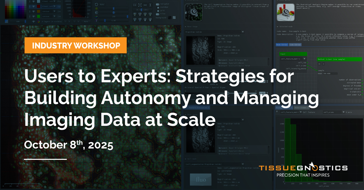
Exposé: from Data to Insights
Alain Pitiot, Senior Project Manager @ TissueGnostics
Dr. Alain Pitiot introduces Exposé, TissueGnostics’ integrated platform for the acquisition, management, visualisation and analysis of biological data. The talk will focus on how Exposé streamlines end-to-end workflows, linking slide scanning, statistical characterization, and visualization within a cohesive, GPU-accelerated environment, and showcase the platform’s flexibility and interoperability with OMERO. Use cases include CellPose integration and dynamic reporting with Exposé dynamic notebooks.
Cancer cell MHC-II expression promotes tumor progression in early lung adenocarcinoma
Francis Jacob Kassama, PhD student at Jacks lab @ MIT
Preliminary data from Jacks lab demonstrate that genetic ablation of cancer cell specific MHC-II expression led to reduced tumor burdens, revealing a novel role of cancer cell MHC-II in influencing CD4+ T cells to help support tumor development. StrataQuest, image analysis software, was employed to examine immune infiltration dynamics in the lung tumor models, enabling visualization and quantification of cellular infiltrates and providing spatial insights.
What you will learn
- Learn from Real-World Experience:
Hear how Dr. Jeffrey Kuhn (MIT) supports over 200 users with a scalable whole slide imaging workflow in a high-demand core setting. - Support Multi-User Environments with Confidence:
Explore practical strategies for managing shared slide scanning systems that promote user independence and operational efficiency. - Simplify Complex Image Analysis:
See how modern analytical platforms enable flexible, whole-slide workflows, easing the generation of reproducible, publication-ready results. - Streamline Imaging Pipelines:
Discover how integrated acquisition and analysis tools can improve throughput while maintaining high data and image quality. - Take Home Actionable Insights:
Gain practical ideas for purchasing, training, and workflow design—relevant to microscopy cores, pathology, immunology, and cancer research. - Engage with Experienced Users:
Connect with peers and solution specialists to ask questions, share challenges, and explore collaborative approaches to imaging and analysis.