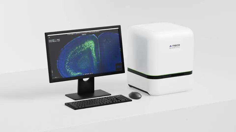
The Atlas Mapped Brain Slice Scanning
PanoBrain™ is an automated platform for brain slice imaging and atlas mapping, designed to accelerate high-throughput neuroscience workflows. Developed by Meca Scientific, PanoBrain transforms prepared tissue sections into high-resolution, atlas-registered digital datasets with minimal manual intervention—bridging the gap between bench and analysis.
- Automated panoramic imaging that preserves full anatomical context across entire tissue sections
- Reproducible, high-throughput acquisition with integrated atlas mapping for large-scale studies
- Analytical workflows that eliminate manual stitching and region-of-interest bias
- Seamless integration with existing histology pipelines and third-party analysis tools
ScientiaLux has more than 15 years of experience working in high-resolution automated slide scanning. Contact us to learn how PanoBrain can power your brain slice imaging and analysis workflows. We’ve got ideas we want to share!
PanoBrain™ Capabilities & Technical Highlights
Automated Imaging & Analysis Pipeline
- Fully automated transition from brain slices to quantitative digital datasets
- Integrated hardware and software pipeline minimizes operator intervention and variability
- Designed to support batch processing of large slide sets
AI-Assisted Brain Mapping & Quantification
- Automated brain region segmentation and atlas registration
- Compatibility with standard reference frameworks (e.g., Allen Brain Atlas)
- Built-in tools for automated cell counting and fluorescence intensity quantification
High-Speed, Large-Format Imaging
- Widefield scanning optimized for rapid, whole-slice acquisition
- Supports large-format slides (up to 125 × 50 mm), enabling full imaging of large brain sections
- Precision XYZ motion platform (0.1 µm) for accurate, repeatable positioning
Multi-Modal & Multi-Color Imaging
- Brightfield and multi-channel fluorescence imaging modes
- Supports complex, multi-label experiments for detailed cellular and circuit-level analysis
Optical & Illumination Performance
- Sub-micron resolution suitable for identifying individual neurons and fine structures
- Highly uniform illumination across the full field of view for seamless panoramic images
- Low-phototoxicity imaging enabled by exposure-synchronized illumination control
Research Applications
PanoBrain is well-suited for applications including:
- Whole-brain and regional circuit mapping
- Viral tracing and connectivity studies
- Quantitative pathology and neurodegeneration research
- Drug efficacy and regional response analysis